

Amyloid plaque detection in the brain with silver staining (NDT403a):
Amyloid plaques can be detected on free-floating sections (e.g. those cut with a cryostat or Vibratome®using FD NeuroSilver™ Kit (cf. Products, Cat. #NDT103), or on sections that have previously been mounted on slides (e.g. paraffin sections) with Hirano-Zimmerman Method¹.
Option 1: Detection of amyloid plaques on free-floating sections with FD NeuroSilver™ Kit
This service includes tissue preparation, sectioning, silver-staining, mounting, coverslipping and labeling the slides. As a result, your will receive up to 40 silver-stained sections per brain or per tissue block ready for microscopic observations. A set of Nissl (or H&E) stained sections adjacent to those used for silver-staining can also be provided at a low cost.

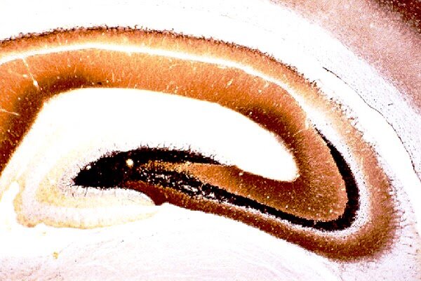
Detection of neurodegeneration and amyloid plaques in the mouse brain of a model for Alzeimer's disease. 40 um cryostat section was cut coronally through the dorsal hippocampus of a transgenic mouse used as a model for studing Alzeimer¡¦s disease. The section was processed for detecting both neuronal damage and amyloid plaques with NDT 103 Kit. Note silver-stained plaques in both the hippocampus and the cortex

High mignification of the hippocampal CA1 area as shown above. Note that in addition to silver-stained amyloid plaques,numerous degenerating fibers are also present in the corpus callosum.
Option 2: Detection of amyloid plaques on paraffin sections with Hirano Zimmerman Method¹
This service includes section pretreatment, silver-staining, coverslipping and labeling the slides. As a result, your will receive up to 40 silver-stained sections per brain or per tissue block ready for microscopic observations. A set of Nissl (or H&E) stained sections adjacent to those used for silver-staining can also be provided at a low cost.
Remarks:
A quotation is required before placing an order.
The investigator needs to provide tissue fixed with formaldehyde or slides with paraffin embedded tissue sections.
Please contact us for more information.
Tissue preparation & Golgi-Cox Staining with FD Rapid GolgiStain (NDT403b):
Golgi-Cox impregnation1, 2 has been one of the most effective techniques for studying both the normal and abnormal morphology of neurons, as well as glia. Using Golgi technique, subtle morphological alterations in neuronal dendrites and dendritic spines have been discovered in the brains of animals treated with drugs as well as in the postmortem brains of patients with neurological diseases3, 4. However, the complex and time-consuming process of Golgi staining has been a major obstacle to the widespread application of this technique.
FD Rapid GolgiStain™ Kit (Cat. #PNDT104), designed based on the principle of the methods described by Ramón-Moliner2, Glaser and Van der Loos5, has overcome most problems associated with the Golgi-Cox technique. The FD Rapid GolgiStain™ Kit has been tested extensively in the brains from several species of animals, as well as in specimens of the postmortem human brain. This kit has not only significantly simplified and improved the Golgi-Cox technique but has also proven to be extremely sensitive and reliable for demonstrating morphological details of neurons and glia, especially dendritic spines (see samples below).
This service includes tissue preparation, sectioning, staining, coverslipping and slide labeling. As a result, you will receive up to all sections from each brain or block which are Golgi-Cox-impregnated and ready for microscopic observations.
Procedure: For details, see the User Manual for FD Rapid GolgiStain™ Kit (Cat. #NDT104).
Remarks:
A quotation is required before placing an order.
The investigator needs to provide tissue fixed with special fixative provided by FD NeuroTechnologies, Inc.
Please contact us for more information.

Human cortex, stained by NDT104, Rapid Golgistain kit. Note the details of Golgi-impregnated soma and axons (lower right).
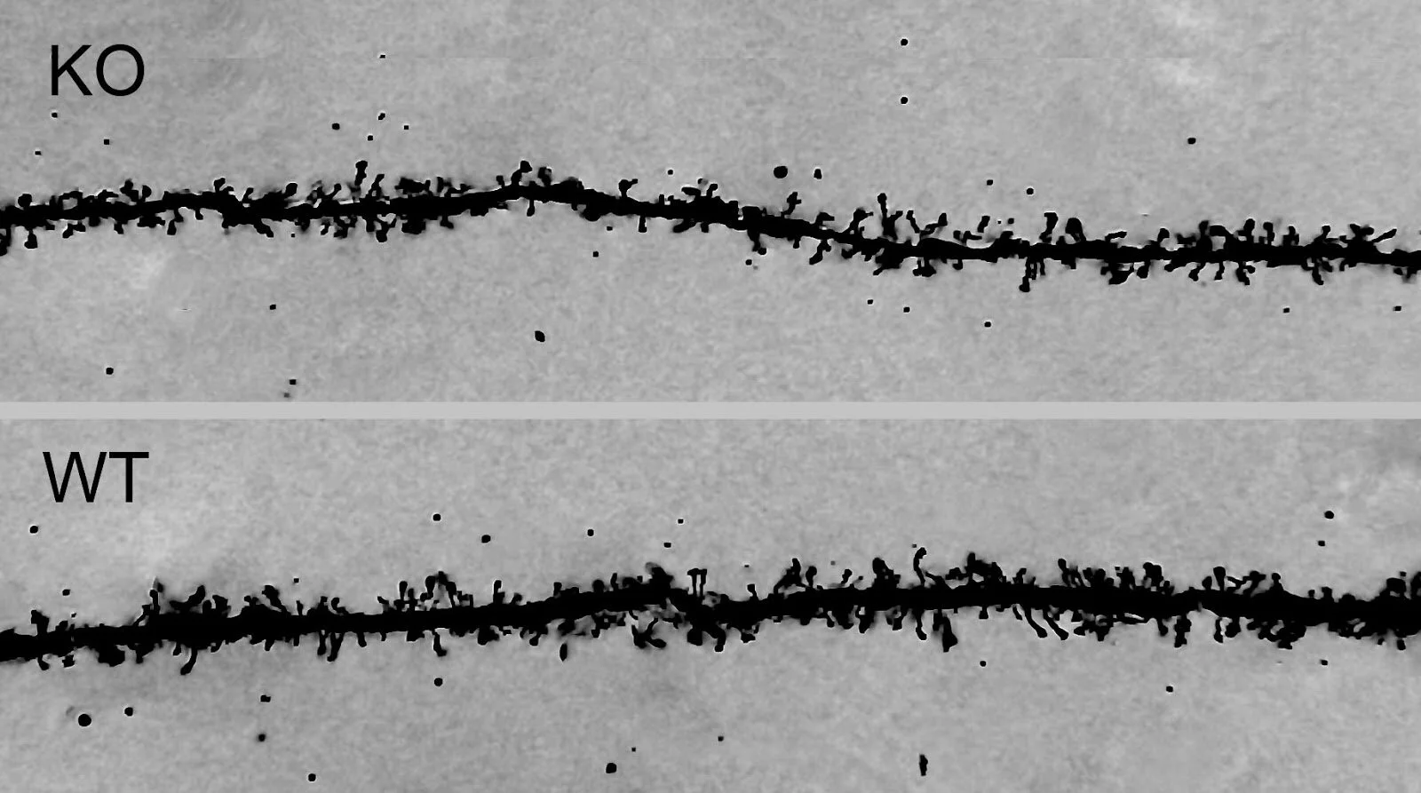
The difference in spine density pf pyramidal cells between KO (knockout) and Wildtype (WT) mice, stained by NDT104, Rapd GolgiStain kit.
Procedure: For details, see the User Manual for FD Rapid GolgiStain™ Kit (Cat. #NDT104).
Remarks:
A quotation is required before placing an order.
The investigator needs to provide tissue fixed with special fixative provided by FD NeuroTechnologies, Inc.
Please contact us for more information.
Timm's Sulphide Silver Staining (NDT403c):
Timm's sulfide silver staining has been used to visualize a variety of metals in brains and other tissues1. Among these are the trace metals essential for life, such as Zn, Cu, Fe, Co, and Ni, as well as toxic metals, e.g., Hg, Cd, Pb, As, Bi, TI, Au and Ag. This method, originally developed by Timm2, was later modified3. The principal of the technique is based on sulphide-precipitation of metals in tissue followed by a physical development. During the latter stage the metal sulphides catalyze the reduction of silver ions by reducing agents. This technique has proven to be particularly useful in visualizing zinc-containing neurons and the detection of newly sprouted axons and axon terminals within the central nervous system (see below).
This service includes tissue preparation, sectioning, staining, coverslipping and labeling the slides. As a result, your will receive up to 40 Timm's sulfide silver stained sections per brain or per tissue block ready for microscopic observations. To express our appreciation, a set of Nissl (or H & E) stained sections adjacent to those used for silver-staining will also be provided for you without additional cost.
Timm's staining of the rat hippocampus. 30 µm cryostat section cut through the dorsal hippocampus of a normal rat was processed for Timm¡¦s histochemistry. Note that the highest density of reaction product is present in mossy fibers traveling in the polymorphic layer of the dentate gyrus and the stratum lucidum of CA3.

Timm's staining in the dentate gyrus of a normal rat. High magnification of the polymorphic, granule cell and molecular layers of the dentate gyrus from the same section as shown above . Note little reaction deposits in both the granule cell and molecular layers.

The seizure-induced morphological changes of the mouse dentate gyrus, countered stained by NDT106 Rapid TimmStain kit and Nissl.

Timm’s staining in the dentate gyrus of a rat model for epilepsy. High magnification of the dentate gyrus from a rat that had developed chronic seizures. Note a dense plexus of reaction product in the inner (supragranular) layer of the dentate gyrus.
NADPH histochemistry of the brain (NDT403d):
Please contact us for more information.
X-Gal histochemistry (NDT403e):
The β-galactosidase gene is often used as a reporter gene, “marker” that is widely used for analysis of mutationally altered genes, as well as gene regulation. The expression of β-galactosidase gene can be detected by histochemical staining of tissue sections or cells. The method is based upon the ability of β-galactosidase to hydrolyze 5-bromo-4-chloro-3-indolyl-β-D-galactopyranoside (X-Gal), resulting in a blue staining in the cells expressing β-galactosidase¹.
This service includes tissue preparation, sectioning, histochemical staining, counterstaining (optional), and coverslipping. As a result, your will receive X-Gal stained sections ready for microscopic observations.
Remarks:
A quotation is required before placing an order.
The investigator needs to provide unfixed and freshly frozen tissue.
Please contact us for more information.
Reference:
Horwitz JP, Chua J, Curby RJ, Tomson AJ, Darooge MA, Fsher be, Mauricio J, Klundt I. (1964) Substrates for cytochemical demonstration of enzyme activity. I. Some substituted 3-Indolyl-Beta-D-Glycopyranosides. J. Med. Chem. 7:574.
Terms and Conditions
For quality assurance of our service, it is recommended that you discuss with us for preferred perfusion protocol and histology and/or immunolabeling protocols.
It is suggested that you use Gel-coated microscopic slides for tissue mounting and 0.17um-thick coverslips.
A 15% of the fee will be due upon authorization of the study; and the remaining fee will be due upon delivery of study results.
Progress of the service is contingent upon staining quality of tissues, operated by the independent contractor.
Should early termination occur, Neurodigitech will prorate the cost incurred and invoice the Sponsor. The first portion of the fee is non-refundable.