
GolgiChromeTM staining Service (Double-stainiing of Golgi impregnation and immunohistochemistry) [Discontinued]
This invention is designed to achieve the simultaneous visualization of Golgi-Cox and immunofluorescence of individual neurons using Confocal Laser Scanning Microscopy (CLSM).
The main advantages of this method are as follows:
Highest visualization of structural details along with the neurochemical nature of the neuron;
Characterization of antigens with specific antibodies
Anatomical interactions among neuronal elements;
The possibility of using confocal laser scanning microscope (CLSM) and accordingly 3D reconstructions and modeling;
Savings of laboratory animals, when up to now it was necessary to perform the two techniques separately and thus on different animals.
Consequent substantial advantages in terms of time and cost saving, together with a higher quality than traditional techniques.
The following examples (Spiga et al., 2012) demonstrate the co-localizations of Golgi-cox impregnated neurons immunoreactive to Tyrosine Hydroxylase (TH) (Figure 1), TH and Postsynaptic Density 95 (PSD-95) (Figure 2), TH, PSD-95 or Synapsin (SynI) (Figure 3), and TH, PSD-95 or SynI of a fully impregnated pyramidal cell (Figure 4).
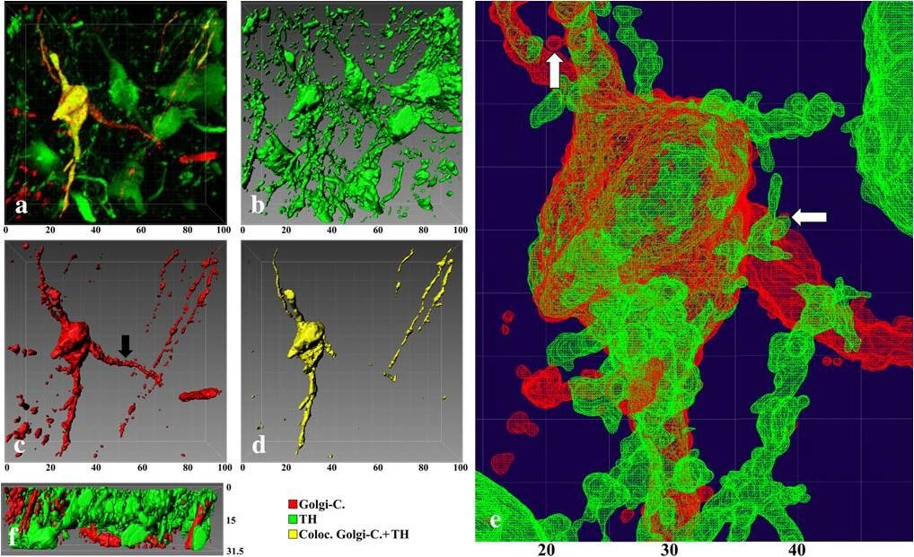
Figure 1. Golgi + Tyrosine hydroxylase (TH) immunohistochemistry in the striatum.

Figure 2. Golgi + TH + PSD95 immunohistochemistry in the substantia nigra (SN, upper panel) and anterior commissure (ACA, lower panel).

Figure 3. Golgi + TH + PSD95 or Synapsin (SynI) immunohistochemistry in the dendritic segments of the medial spiny neurons (MSN) of the striatum.

Figure 4. Golgi + TH + PSD95 or SynI immunohistochemistry of the prefrontal prelimbic zone of the rat brain.
*Analysis of Variance (ANOVA) test will be performed!
Terms and Conditions
For quality assurance of our service, it is recommended that you discuss with us for preferred perfusion protocol and histology and/or immunolabeling protocols.
It is suggested that you use Gel-coated microscopic slides for tissue mounting and 0.17um-thick coverslips.
A 15% of the fee will be due upon authorization of the study; and the remaining fee will be due upon delivery of study results.
Progress of the service is contingent upon staining quality of tissues, operated by the independent contractor.
Should early termination occur, Neurodigitech will prorate the cost incurred and invoice the Sponsor. The first portion of the fee is non-refundable.